Fuch’s Corneal Dystrophy

Voted Best of Berks—
eight years in a row!
Perspective: Although Fuch’s Corneal Dystrophy is a relatively rare type of corneal dystrophy, its impact on patient functioning, comfort and lifestyle can be profound. Medical treatment in the early phases and corneal transplantation in the intermediate and later phases has been fairly effective in helping patients resume normal activities and lifestyles with minimal symptoms. This has been especially true with the increased popularity of advanced corneal transplantation procedures that allow transplantation of the deep corneal layers using “endothelial keratoplasty” or “posterior lamellar keratoplasty” techniques.
Description, Signs & Symptoms: Fuch’s Dystrophy is a slowly progressive disease of the cornea that is typically bilateral and is slightly more common in women than men. While it is possible to observe Fuch’s Dystrophy in people in their 30’s and 40’s, it usually does not compromise vision until people are in their 50’s or 60’s. While a precise path of genetic transmission is unclear, a familial predisposition seems to exist among those who are first degree relatives.
The innermost layer of cells in the cornea, called the endothelium, is a single layer of non-regenerating cells. The endothelial cells are responsible for pumping water out of the cornea and helping to maintain the corneal transparency. While the exact etiology is poorly understood, in Fuch’s Dystrophy, the endothelial cells die, which makes the endothelium less efficient in maintaining the proper level of corneal hydration/dehydration through its normal metabolic pumping activity. This results in the cornea swelling and distorting vision and may be accompanied by disabling glare, night vision problems, haloes and photophobia. In its later phases, Fuch’s Dystrophy is often associated with considerable pain as the epithelium “blisters” by forming “bullae”, which actually begin to rupture.
Early in the course of Fuch’s Dystrophy patients typically awaken with blurry vision that gets progressively clearer as the day passes. This phenomenon occurs because the cornea normally swells during sleep. A healthy endothelial pump is necessary in order to restore the cornea to a normal thickness. In Fuch’s Dystrophy, the endothelium removes fluid from the cornea less efficiently. As the Fuch’s Dystrophy disease worsens, the vision does not clear. Usually we will attempt to help reduce the corneal swelling each day by prescribing hypertonic eye drops and ointments. However, when these measures fail to provide comfort and clear vision, it may be necessary to have a corneal transplant.
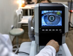
Testing & Diagnosis: The diagnostic exam for Fuch’s Dystrophy will generally include at a minimum,
a) visual acuity measurement
b) glare testing to determine whether visual acuity degradation occurs under bright light conditions-this often correlates with subjective complaints of night driving difficulty
c) evaluation of the presence and grade of “guttata”-using the slit lamp biomicroscope to examine the corneal endothelium we can assign a “grade” or “stage” of zero through five to the presence of guttata to indicate the severity of dystrophy with zero indicating no disease and five indicating severe disease.
d) endothelial cell count-using the specular microscope we are able to obtain both a cell count and morphological evaluation within a specific area of the cornea; low cell counts and morphological changes-polymegathesim and pleomorphism-indicate the presence of disease.
e) tonometry for intraocular pressure measurement
f) pachymetry, typically performed by ultrasonic or optical methods, is used to determine corneal thickness; increased thickness indicates edema and thus inefficient endothelial function.
Symptoms
- blurry vision on awakening that may gradually clear as the day progresses
- pain from epithelial bullae on the corneal surface
- overall visual impairment, distorted vision and reduced contrast
- poor night vision
- photophobia
- glare
- haloes
About Cornea Transplants for Fuch’s Dystrophy: In general, we use corneal transplantation in order to replace diseased, damaged or scarred corneal tissue with new healthy corneal tissue. Since damaged or scarred corneal tissue impairs effective passage of light into the eye and may distort of degrade the quality of images that reach the retina, poor vision and even blindness may result from a damaged cornea. There are actually a number of different types of corneal transplants that we can consider for Fuch’s Dystrophy:
Penetrating Keratoplasty (PK): This type of corneal transplant involves the surgical removal of the central two-thirds thickness of the damaged cornea. We remove the central portion of the damaged or cloudy cornea with a “cookie cutter” like instrument called a trephine and replace it with a “button” of clear cornea obtained from the eye bank. We then carefully suture the donor cornea button into place. We typically will use ___sutures and work to attempt to control any induced astigmatism. Once the new corneal tissue has healed properly, we can remove the sutures. It is not uncommon to have some degree of induced refractive error depending on the effective suture tension and tissue healing pattern. This may require eyeglass and/or contact lens correction in some cases. In many cases we are able to use the excimer laser to achieve the final correction with Photorefractive Keratectomy. This type of transplant has the potential to provide the clearest vision after healing because there is no interface (layer) to look through. However, the healing time is somewhat long.
Decemet’s Stripping with Endothelial Keratoplasty (DSEK): For some patients, this a relatively new procedure, called DSEK (Decemet’s Stripping with Endothelial Keratoplasty) allows us to transplant endothelial cells, which may help certain Fuch’s Dystrophy patients overcome their discomfort and vision problem.
We perform DSEK through a small incision in order to remove and replace the cornea endothelial layer. First we gently “strip” off the single diseased endothelial cell layer and leave the remaining cornea intact. Next, we will thinly slice a donor cornea from the eye bank and fold the back portion in half and insert it through the small incision into the eye. By using an injected air bubble we are able to unfold and position the donor tissue on the recipient cornea. Within a few minutes the donor tissue attaches to the recipient without the use of any sutures. There are a number of advantages of DSEK for patients who are deemed to be good candidates:
- better structural integrity of the eye itself
- rapid visual recover
- little if any change in refractive status of the eye
Cornea transplants have become somewhat common in the United States as a treatment for damaged and cloudy corneas. Each year more that 40,000 people undergo corneal transplantation to restore their vision. Cornea transplants have proven to be quite effective for Fuch’s Dystrophy. In fact, the majority of patients who have a successful transplant for Fuchs’ Dystrophy continue to be free of symptoms for at least ten years. With the increasing popularity of advanced cornea transplantation utilizing endothelial keratoplasty and posterior lamellar keratoplasty techniques such as DSEK (Decemet’s Stripping with Endothelial Keratoplasty), we are able to treat the visual debilitation and discomfort of Fuch’s Dystrophy with even greater confidence.
Please feel free to contact us regarding information for patients with Fuch’s Dystrophy or any corneal disease or problem. Drs. Altman, Primack, and Shah are Fellowship trained Cornea Specialists at Eye Consultants of Pennsylvania, P.C. and may be reached at 610-378-1344.
Find a Doctor
Physician information including education, training, practice location and more.
Schedule an Appointment
Call 800-762-7132 or make an appointment online.