Learn more about macular degeneration
What is macular degeneration? Symptoms, treatment options and more!

At a Glance:
Things to know and remember:
- There are two forms of age-related macular degeneration -- dry and wet.
- The risk for age-related macular degeneration (AMD) increases with age. AMD is most common in older people, but it can occur during middle age.
- Other risk factors include smoking, obesity, race, family history and gender.
- AMD causes no pain, but an early symptom of wet AMD is that straight lines appear wavy. The most common symptom of dry AMD is slightly blurred vision.
- Vision lost cannot be restored. That’s why early detection and treatment are important.
About Macular Degeneration
What is macular degeneration?
Macular degeneration, or age-related macular degeneration (AMD) is a leading cause of vision loss in Americans 60 and older. It is a disease that destroys your sharp, central vision. You need central vision to see objects clearly and to do tasks such as reading and driving.
AMD affects the macula, the part of the eye that allows you to see fine detail. It does not hurt, but it causes cells in the macula to die. In some cases, AMD advances so slowly that people notice little change in their vision. In others, the disease progresses faster and may lead to a loss of vision in both eyes. Regular comprehensive eye exams can detect macular degeneration before the disease causes vision loss. Treatment can slow vision loss. It does not restore vision.
Diagnosis & Treatment Options
How is AMD diagnosed?
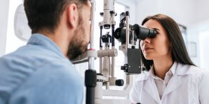
AMD is detected during a comprehensive eye exam that includes a multitude of tests. Tests for AMD include:
- The visual acuity test is an eye chart test that measures how well you see at various distances.
- In the dilated eye exam, drops are placed in your eyes to widen, or dilate, the pupils. Then, your eye care professional uses a special magnifying lens to examine your retina and optic nerve for signs of AMD and other eye problems. After the exam, your close-up vision may remain blurred for several hours.
- With tonometry, an instrument measures the pressure inside the eye. Numbing drops may be applied to your eye for this test.
Your eye care professional also may do other tests to learn more about the structure and health of your eye.
 The Amsler Grid: During an eye exam, you may be asked to look at an Amsler grid, shown here. You will cover one eye and stare at a black dot in the center of the grid.
While staring at the dot, you may notice that the straight lines in the pattern appear wavy. You may notice that some of the lines are missing. These may be signs of AMD.
Because dry AMD can turn into wet AMD at any time, you should get an Amsler grid from your eye care professional. You could then use the grid every day to evaluate your vision for signs of wet AMD.
The Fluorescein Angiogram Test: If your eye care professional believes you need treatment for wet AMD, he or she may suggest a fluorescein angiogram. In this test, a special dye is injected into your arm. Pictures are taken as the dye passes through the blood vessels in your eye. The test allows your eye care professional to identify any leaking blood vessels and recommend treatment.
OCT: A procedure to measure retinal thickness.
Does my insurance plan
cover my eye care?
Find out what insurance we accept and what is covered by insurance.
Learn more about our macular degeneration specialists
Physician information including education, training, practice location and more.
Schedule an Appointment
Schedule an appointment with one of our specialists.