Keratoconus

Voted Best of Berks—
eight years in a row!
Perspective: Although Keratoconus is a relatively rare corneal disease, its impact on vision and overall lifestyle can be quite significant as it progresses. Vision correction in the early phases is often possible with eyeglasses and contact lenses. In the intermediate phases specialized contact lenses are often necessary to provide good vision and physical tolerance. As Keratoconus progresses and reaches its later phases, corneal surgery including transplantation may be required in order for patients to conduct normal activities and enjoy normal lifestyles. Newer types of corneal surgery and treatment may include implantation of prescription inserts, called Intacs®, and a non surgical approach called Holcomb C-3R® CXL that enhances corneal collagen cross linking through the photoactivation of riboflavin. By carefully monitoring the progression of Keratoconus and applying the necessary treatment options it is possible to help patients maintain good vision and functioning.
Description, Signs & Symptoms: Keratoconus is a non inflammatory “ectasia” of the cornea in which the normally round dome-shaped cornea progressively thins causing a cone-like bulge to develop resulting in significant visual impairment. The actual incidence of Keratoconus is unknown. However, Keratoconus has been estimated to occur in 1 out of every 1-2,000 persons in the general population. Keratoconus is generally first diagnosed in young people at puberty or in their late teens and progresses through the third or fourth decade of life. It tends to progress more rapidly in young patients. Keratoconus occurs about equally in men and women. It is found in all parts of the United States and the rest of the world. It has no known significant geographic, cultural or social pattern. The signs and symptoms of Keratoconus may change as the disease progresses and may include a) blurred or distorted vision b) monocular double vision c) rapidly changing eyeglass prescriptions especially with high degrees of astigmatism d) increased sensitivity to bright light and glare e) problems with night vision f) headaches from eyestrain and in the most severe situation e) hydrops, a painful condition in which the back of your cornea ruptures and fills with fluid, causing a sudden clouding and loss of vision.
Keratoconus is often an isolated ocular condition. However, sometimes it is found to exist in conjunction with eye and systemic conditions. Some of the more common conditions that we see along with Keratoconus include atopic dermatitis, vernal conjunctivitis and connective tissue disorders including Marfan’s and Ehlers-Danlos Syndrome as well as mitral valve prolapse. There is the belief that several factors may actually predispose you to developing Keratoconus. These may include:
Wearing Poorly Fit Rigid Contact Lenses-If you wear rigid contact lenses and they haven’t been fitted properly, the constant pressure or continual injury they cause can lead to Keratoconus.
Certain Inherited Diseases-The risk of developing Keratoconus is higher if you have certain conditions, such as Down Syndrome or certain retinal diseases such as Retinitis Pigmentosa.
Family History of Keratoconus-Most people with Keratoconus have no family history of the disease, but researchers have found that in some cases there may be a family connection.
While most cases of Keratoconus are not preventable it is still worthwhile taking some simple precautionary steps to be sure you don’t cause it yourself:
Use Care When Rubbing Your Eyes. If you vigorously rub your eyes daily, you may be causing cumulative damage to your corneas in a way that can ultimately lead to Keratoconus. Some people don’t realize the extent of their eye-rubbing habit until a family member or friend points it out, so pay attention if someone has mentioned it. Try to wipe or rub your eyes gently and as little as possible.
Follow Instructions When Wearing Rigid Contact Lenses. If you wear rigid contact lenses, be sure they’ve been fitted correctly and use them as directed to avoid damage to your eyes. Never use someone else’s hard contact lenses.
Management & Treatment of Keratoconus:
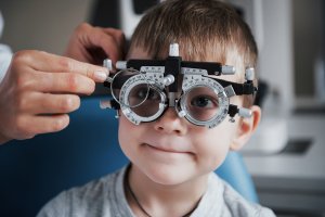
Eyeglasses and soft contact lenses may be well tolerated and provide satisfactory comfort and vision when Keratoconus is mild. Most often, the quality of vision tends to decline so that patients require Rigid Gas Permeable Lenses (RGP) for vision correction. Other contact lens options for Keratoconus include piggyback lens systems where an RGP lens in worn over a soft silicone hydrogel lens, hybrid contact lenses such as SoftPerm® and SynergEyes® and even very large gas permeable contact lenses that fit over the sclera or white of the eye. When good contact lens care is available only 10-20% of patients progress to the point of needing medical or surgical intervention.
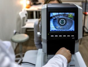
Collagen Cross Linking-Holcomb C-3R® CXL is a non-invasive treatment that has been proven to strengthen the weak corneal structure in Keratoconus. This treatment method works by increasing collagen cross-linking, which serve as the natural “anchors” within the cornea. These anchors are responsible for preventing the cornea from bulging and becoming steep and irregular and thus causing Keratoconus. Holcomb C-3R® CXL is administered by placing riboflavin eye drops onto the surface of the eye and exposing the surface to a precise amount of ultraviolet light. When this method receives final Food and Drug Administration (FDA) approval it is expected to offer a possible solution for many Keratoconus patients who have progressed to a significant amount of visual disturbance but not so much so as to require a cornea transplant.
Intacs™ Prescription Inserts-Intacs™ Prescription Inserts for Keratoconus are useful as an intermediate treatment between contact lenses and a corneal transplant. Intacs™ prescription inserts may help to stabilize the cornea and improve vision. These prescription inserts are indicated for use in the correction of nearsightedness and astigmatism for patients with Keratoconus, where contact lenses and glasses are no longer suitable. They are inserted into microscopic channels created in the cornea by a corneal surgeon. Intacs™ inserts may be combined with C-3R® CXL if additional structural stability is required in the cornea, but not so much as to require a cornea transplant.
Corneal Transplantation-When Keratoconus has progressed to the point that contact lenses are no longer well tolerated and C-3R® CXL with or without Intacs™ can not provide good vision correction, a cornea transplant is the best choice. In most cases of Keratoconus a penetrating keratoplasty (PKP) is the preferred surgical solution although sometimes the cornea surgeon may recommend a lamellar keratoplasty (LKP). With PKP the cornea surgeon removes the central portion of the damaged cornea with a “cookie cutter” like instrument called a trephine and replaces it with a clear cornea obtained from the eye bank. The donor cornea is very carefully sewn into place using sutures that are thinner than a human hair. When the new cornea has healed properly, the fine sutures or stitches can be removed right in the office in most cases. PKP is the most common type of cornea transplant as it has the potential to provide the clearest vision after healing because there is no interface (layer) to look through. However, the healing time is somewhat long and the use of a contact lens might be required for the clearest vision. Sometimes Lamellar Keratoplasty (LKP) may be used if the damaged corneal tissue is mainly located in the outermost 50% of the cornea. In LKP the outermost half of the cornea is carefully dissected and removed along with any damaged tissue. Then a new donor cornea is gently sewn into place. This type of corneal transplant is less invasive and will allow your eye to be stronger after surgery than it would be with a regular full thickness transplant, or Penetrating Keratoplasty. However, in some cases there can be some loss of clarity from the interface between the new and remaining layers of the cornea.
Symptoms
- blurred or distorted vision
- monocular diplopia or shadowing
- large increases in prescription, especially astigmatism and myopia
- increased sensitivity to glare
- photophobia
- headaches
- sudden painful clouding and loss of vision
For patients with virtually any degree of Keratoconus we are now able to offer them a range of treatment options that will allow for a good long term prognosis and vision correction. We are able to treat the visual debilitation and discomfort of Keratoconus with more confidence than ever.
Please feel free to contact us regarding information for patients with Fuch’s Dystrophy or any corneal disease or problem. Drs. Altman, Primack, and Shah are Fellowship trained Cornea Specialists at Eye Consultants of Pennsylvania, P.C. and may be reached at 610-378-1344.
Find a Doctor
Physician information including education, training, practice location and more.
Schedule an Appointment
Call 800-762-7132 or make an appointment online.