Tear Duct Surgery

Voted Best of Berks—
eight years in a row!
Tearing in Adults
Excess tearing in adults may be caused either by poor tear drainage or an overproduction of tears.
Tears are produced in the tear gland. By lubricating the eyes, tears play a vital role in the maintaining the health of the eye. When the eye is irritated excess tears are produced. People are not always aware of the eye irritation, but instead may notice the overproduction of tears.
Tears normally drain into small holes located in the inside corner of the upper and lower eyelids. The tears then collect in the tear sac. The tear sac lies under the skin between the corner of the eye and the nose. Next, the tears flow through a small tube, called the nasolacrimal duct, into the nose. The tears are pumped through this drainage system by the opening and closing of the eye.
A blockage in any part of this drainage system can prevent tear drainage and lead to excess tears running out of the eye and down the cheek.
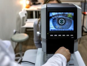
It is very important to properly diagnose which part of the tear drainage system is blocked in order to determine the proper treatment. A harmless orange dye will be placed in the eye in order to see if the tears drain normally. A thin metal tube will then be placed into the tear drainage system in order to see if the tubes leading into the tear sac are open. The doctor then will attempt to flush water through the nasolacrimal duct into the nose in order to determine if the nasolacrimal duct is open.
In some patients excess tearing is caused by several different causes. Patients may have a partial blockage of the tear drainage system, poor pumping of tears because of eyelid weakness and excess tear production due to eye irritation, all at the same time.
Treatment of Tear System Blockage
- When the nasolacrimal duct, the tube which drains tears into the nose, is blocked a surgical procedure is usually required. During this procedure, called a DCR (Dacryocystorhinostomy), a hole is created between the tear sac and the inside of the nose.
- When the nasolacrimal duct is only partially blocked your doctor will often attempt to widen the opening by flushing water through the duct. An eyedrop with both anti-inflammatory and antibiotic medications will then be prescribed in an attempt to reduce the swelling in the duct and promote tear drainage. These attempts to open the duct are often successful but may need to be repeated periodically
- When the tubes, called cannaliculli, which drain the tears into the lacrimal sac are blocked surgical therapy is more complicated. In most cases both a DCR and reconstruction of the cannaliculli will be performed.
- Eyelid weakness or malposition may be treated by surgically tightening and repositioning the lids.
DCR – Surgical Procedure
DCR is a same-day procedure. You may choose to be either asleep under general anesthesia or awake with sedation.
A DCR is performed through a skin incision, which is made on the side of the nose. The bone between the tear sac and the nose is removed, and the lining of the tear sac is then attached to the lining of the nose to form a permanent drainage for tears.
In some people, during surgery, a clear plastic tube is placed from the inside corner of the eye into the nose. The tube is used to stent the tear drainage system and prevent scarring. This tube is easily removed in the office in two months.
Sutures are placed in the skin and removed in about a week. The scar produced will initially be red, hard and raised but will smooth out over the months after surgery.
Options
Various other procedures have been attempted in order to open a completely blocked nasolacrimal duct. However, these attempts often do not work in the long term. Therefore, at this time, Dr. Goldberg believes a DCR is the best option for most people.
A DCR can also be performed through the nose (called an endoscopic DCR). This avoids a skin incision. However, the success rate with this procedure is probably less than when the procedure is performed through the skin because the tear sac is not directly attached to the lining of the inside of the nose.
Please feel free to discuss these options with Dr. Goldberg.
Problems
Most people are very satisfied with the results of their tear duct surgery. However, in about 10 – 20% of people excess tearing will still be present after surgery or will recur. There are three common reasons for surgical failure.
- Scarring of the hole between the lacrimal sac and the nose – This is probably the most common cause of surgical failure. Scarring may occur soon after surgery or years later. If scarring occurs, the drainage hole can often be opened in the office. In some people, the hole must be widened in the hospital either through the nose or through a skin incision.
- Scarring of the drainage tubes (cannaliculli) leading into the tear sac – This is a more difficult situation to treat. In some cases plastic tubes can be placed through the natural drainage tubes to keep them open.
- Poor eye pumping – Some people may have excess tearing even if the tear drainage system produced by surgery is wide open. In this case various eyelid procedures may be attempted.
Other complications of a DCR include an unsatisfactory skin scar and infection. A few people have severe nose bleeds after surgery which may require treatment by an Ear, Nose & Throat specialist.
INSTRUCTIONS BEFORE SURGERY
Please see the separate Instructions Before Hospital Surgery information.
INSTRUCTIONS AFTER SURGERY
Prevention of nose bleeds
In order to prevent nasal bleeding, please do not blow your nose for five days after surgery. It is also important to keep the inside lining of your nose moist. If your house is dry, a humidifier is recommended.
Only light activity should be performed the first two days. Any blood pressure medicine you are taking should be continued.
There may be some bloody nasal discharge for a few days after surgery. If mild nasal bleeding occurs it can be stopped by pinching your nostrils closed and sitting upright for 10 minutes. If bleeding continues, please call our office or go to the Emergency Room.
Afrin or ordinary saline nose drops may be used to clear the mucous discharge from the nostril.
Cold Compresses
In order to reduce swelling, please apply ice packs or cold compresses to the incision at least four times per day for the first two days following surgery.
Medication
After surgery you should continue the medications you normally take, especially blood pressure medications. The use of blood thinners such as coumadin and aspirin should be discussed individually with Dr. Goldberg.
You will be asked to use an antibiotic ointment on your wound three times per day beginning the day after surgery. The antibiotic ointment should be continued until the sutures are removed.
An antibiotic / anti-inflammatory eyedrop should also be placed in the operated eye three times per day for two weeks.
If pus is found in the tear sac during surgery you will also be placed on an oral antibiotic.
Wound Care
A dressing will be placed on the wound. This dressing can be removed the morning after surgery. A gauze may also be taped under your nose to catch any nasal discharge. This gauze may be removed when you get home from surgery or sooner if desired.
You should not work in the yard, get the wound dirty, or go swimming for two weeks following surgery. However, you may wash your hair, bathe, or go to the hairdresser beginning the day after surgery. If dried blood and discharge builds up on the wound you should clean the wound with peroxide on a Q-tip.
If you notice pus, fever, or marked redness and tenderness of the wound you may have an infection. Please call our office.
Not every condition is listed. Consult Dr. Goldberg if you have any questions about a condition not described here.
Find a Doctor
Physician information including education, training, practice location and more.
Schedule an Appointment
Call 800-762-7132 or make an appointment online.