Glaucoma

Voted Best of Berks—
eight years in a row!
What is GLAUCOMA?
Glaucoma is a group of eye diseases characterized by damage to the optic nerve, the nerve that transmits visual information from the eye to the brain. In most cases, this nerve damage is produced by increased fluid pressure within the eye. This elevated pressure is caused by a backup of fluid in the eye. Over time, it causes damage to the optic nerve. Through early detection, diagnosis and treatment, you and your doctor can help to preserve your vision.
Think of your eye as a sink, in which the faucet is always running and the drain is always open. The aqueous humor is constantly circulating through the anterior chamber. It is produced by a tiny gland, called the ciliary body, situated behind the iris. It flows between the iris and the lens and, after nourishing the cornea and lens, flows out through a very tiny spongy tissue, only one-fiftieth of an inch wide, called the trabecular meshwork, which serves as the drain of the eye. The trabecular meshwork is situated in the angle where the iris and cornea meet. When this drain becomes clogged, aqueous cannot leave the eye as fast as it is produced, causing the fluid to back up. But since the eye is a closed compartment, your `sink´ doesn´t overflow; instead the backed up fluid causes increased pressure to build up within the eye. We call this open (wide) angle glaucoma.
To understand how this increased pressure affects the eye, think of your eye as a balloon. When too much air is blown into the balloon, the pressure builds, causing it to pop. But the eye is too strong to pop. Instead, it gives at the weakest point, which is the site in the sclera at which the optic nerve leaves the eye.
The optic nerve is part of the central nervous system and carries visual information from the eye to the brain. This cranial nerve is made up of over one million nerve axons, which are nerve fiber extensions of the retinal ganglion cells. When the eye pressure is increased and/or other inciting factors exist, the optic nerve becomes damaged and the retinal ganglion cells undergo a slow process of cell death termed “apoptosis.” The death of the retinal cells and degeneration of the nerve fibers results in permanent vision loss. Early diagnosis and treatment of glaucoma can help prevent blindness.
Who is at risk for Glaucoma?
Everyone should be concerned about glaucoma and its effects. It is important for each of us, from infants to senior citizens, to have our eyes checked regularly, because early detection and treatment of glaucoma are the only way to prevent vision impairment and blindness. There are a few factors related to this disease which tend to put some people at greater risk:
People over the age of 40: While glaucoma can develop in younger patients, it occurs more frequently as we get older.
People who have a family history of glaucoma: Glaucoma appears to run in families. The tendency for developing glaucoma may be inherited. However, just because someone in your family has glaucoma does not mean that you will necessarily develop the disease.
People with abnormally high intraocular pressure (IOP): High IOP is the most important risk factor for glaucomatous damage.
People of African, Latino, and Asian descent: People with African and Latino ancestry have a greater tendency for developing primary open-angle glaucoma than do people of other races. People of Asian descent are more prone to develop angle-closure glaucoma and normal-tension glaucoma.
People who have:
Diabetes
Myopia (nearsightedness) or Hyperopia (farsightedness)
Regular, long-term steroid/cortisone use
A previous eye injury
History of Sleep Apnea
Extremely high or low blood pressure
Everyone under 40 should have a comprehensive eye examination every three to four years. Individuals under 40 with one of the above risk factors should get tested every one and a half to two years. Everyone 40 years or older should have a comprehensive eye examination every one and a half to two years. If you are 40 and have an additional risk factor listed above, get tested annually. Anyone with high risk factors should be tested every year or two after 35.
What are the different types of Glaucoma?
There are a variety of different types of glaucoma. The most common forms are:
- Primary Open-Angle Glaucoma
- Normal Tension Glaucoma
- Angle-Closure Glaucoma
- Pigmentary Glaucoma
- Exfoliation Syndrome
- Trauma-Related Glaucoma
- Childhood Glaucoma
How is Glaucoma diagnosed?
Your eye doctor has a variety of diagnostic tools that aid in determining whether or not you have glaucoma — even before you have any symptoms. Let us explore these tools and what they do.
The Tonometer: The tonometer measures the pressure in your eye. Your doctor places a numbing eye drop in your eye. Then you sit at a slit-lamp, resting your chin and forehead on a support that keeps your head steady. The lamp, which lets your doctor see a magnified view of your eye, is moved forward until the tonometer, a plastic prism, barely touches the cornea to measure your IOP. The test is quick, easy and painless.
The Pachymeter: The pachymeter measures central corneal thickness (CCT). Like the tonometer, your doctor will first anesthetize your eyes. Then a small probe will be placed perpendicular to the central cornea.
CCT is an important measure and helps your doctor interpret your IOP levels. Some people with thin central corneal thickness will have pressures that are actually higher than when measured by tonometry. Likewise, those with thick CCT will have a true IOP that is lower than that measured. Measuring your central corneal thickness is also important since recent studies have found that thin CCT is a strong predictor of developing glaucoma in patients with high IOP.
Visual Field Test: Visual field is an important measure of the extent of damage to your optic nerve from elevated IOP. In glaucoma, it is the peripheral (side) vision that is most commonly affected first. Testing your visual field lets your doctor know if peripheral vision is being lost. There are several methods of examination available to your doctor; visual field testing has advanced significantly in recent years.
In computerized visual field testing you will be asked to place your chin on a stand, which appears before a concave computerized screen. Whenever you see a flash of light appear, you press a buzzer. At the end of this test, your doctor will receive a printout of your field of vision. New software has been developed to help your doctor analyze these tests as well as monitor progression of visual field loss over successive tests.
Ophthalmoscopy: Using an instrument called an ophthalmoscope, your eye doctor can look directly through the pupil at the optic nerve. Its color and appearance can indicate whether or not damage from glaucoma is present and how extensive it is. This technique remains the most important in diagnosing and monitoring glaucoma.
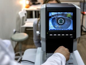
Imaging Technology: A number of new and highly sophisticated image analysis systems are now available to evaluate the optic nerve and retinal nerve fiber layer, the areas of the eye damaged by glaucoma. These devices include scanning laser tomography (e.g. HRT3), laser polarimetry (e.g. GDX), and ocular coherence tomography (e.g. older time-domain OCT or newer spectral-domain OCT). These instruments can help your doctor by giving a quantitative measure of the anatomical structures in the eye. Photographs of the optic nerve can also be useful to follow the progression of damage over time. Large databases have been established to compare an individual’s anatomic structures to those of other patients in the same age group. This software and technology are developing rapidly and show great promise. However, they have not yet evolved to replace ophthalmoscopy, where the doctor looks directly at the optic nerve.
Gonioscopy: Your doctor may perform gonioscopy to closely examine the trabecular meshwork and the angle where fluid drains out of the eye. After numbing the eye with anesthetic drops, the doctor places a special type of hand-held contact lens, with mirrors inside, on the eye. The mirrors enable the doctor to view the interior of the eye from different directions. In this procedure, the doctor can determine whether the angle is open or narrow. Individuals with narrow angles have an increased risk for a sudden closure of the angle, which can cause an acute glaucoma attack. Gonioscopy can also determine if anything, such as abnormal blood vessels or excessive pigment, might be blocking the drainage of the aqueous humor out of the eye.
What are the treatments for Glaucoma?
Glaucoma can be treated with eye drops, pills, laser surgery, traditional surgery or a combination of these methods. The goal of any treatment is to prevent loss of vision, as vision loss from glaucoma is irreversible. The good news is that glaucoma can be managed if detected early, and that with medical and/or surgical treatment, most people with glaucoma will not lose their sight.
Taking medications regularly, as prescribed, is crucial to preventing vision-threatening damage. That is why it is important for you to discuss side effects with your doctor. While every drug has some potential side effects, it is important to note that many patients experience no side effects at all. You and your doctor need to work as a team in the battle against glaucoma. Your doctor has many options. They include:
Eye Drops: It is important to take your medications regularly and exactly as prescribed if you are to control your eye pressure. Typically glaucoma eyedrops are dosed with one drop once or twice a day in the affected eye. Medications should be taken on a fixed schedule every day to achieve the best long term control of the disease. Since eye drops are absorbed into the bloodstream, tell your doctor about all medications you are currently taking. Ask your doctor and/or pharmacist if the medications you are taking together are safe. Some drugs can be dangerous when mixed with other medications. To minimize absorption into the bloodstream and maximize the amount of drug absorbed in the eye, close your eye for one to two minutes after administering the drops and press your index finger lightly against the inferior nasal corner of your eyelid to close the tear duct which drains into the nose. While almost all eye drops may cause an uncomfortable burning or stinging sensation at first, the discomfort should last for only a few seconds.
Oral Medications: Sometimes, when eye drops don’t sufficiently control IOP, pills may be prescribed in addition to drops. These pills (ie. Acetazolamide or Methazolamide) have more systemic side effects than drops. They also serve to turn down the eye’s faucet and lessen the production of fluid. These medications are usually taken from two to four times daily. It is important to share this information with all your other doctors so they can prescribe medications for you that will not cause potentially dangerous interactions.
Surgical Procedures: When medications do not achieve the desired results, or have intolerable side effects, your ophthalmologist may suggest surgery.
Laser Surgery: Laser surgery has become increasingly popular as an initial treatment or an intermediate step between drugs and traditional surgery though the long-term success rates are variable. The most common type performed for open-angle glaucoma is called trabeculoplasty. This procedure takes between 2 and 5 minutes, is painless, and can be performed in either a doctor’s office or an outpatient facility. The laser beam (a high energy light beam) is focused upon the eye’s drain, called the Trabecular Meshwork. Contrary to what many people think, the laser does not burn a hole through the eye. Instead, the eye’s drainage system is changed in very subtle ways so that aqueous fluid is able to pass more easily out of the drain, thus lowering IOP.
You may go home and resume your normal activities following surgery. Your doctor will likely check your IOP one to two hours following laser surgery. After this procedure, many patients respond well enough to be able to avoid or delay surgery. While it may take a few weeks to see the full pressure-lowering effect of this procedure, during which time you may have to continue taking your medications, some patients are eventually able to discontinue some of their medications. This, however, is not true in all cases. Your doctor is the best judge of determining whether or not you will still need medication. Complications from laser are minimal, which is why this procedure has become increasingly popular and some centers are recommending the use of laser before drops in some patients.
Selective Laser Trabeculoplasty (SLT) — for open-angle glaucoma: SLT is a laser that uses very low levels of energy to help stimulate the drainage network to drain more fluid out of the eye and into the bloodstream. It is termed “selective” since it specifically targets portions of the trabecular meshwork. Typically this procedure carries a 75-80% success rate as far as lowering intraocular pressure with very minimal risk. Additionally, SLT may be safely repeated if needed for further control later in life if the disease is noted to progress.
Laser Peripheral Iridotomy (LPI) — for angle-closure glaucoma: This procedure is used to make an opening through the iris, allowing aqueous fluid to flow from behind the iris directly to the anterior chamber of the eye. This allows the fluid to bypass its normal route. LPI is the preferred method for managing a wide variety of angle-closure glaucomas that have some degree of pupillary blockage. This laser is most often used to treat an anatomically narrow angle and prevent both acute and chronic forms of angle-closure glaucoma.
Cycloablation: Two laser procedures for open-angle glaucoma involve reducing the amount of aqueous humor in the eye by destroying or suppressing part of the ciliary body, which produces the fluid.
Transscleral cyclophotocoagulation uses a laser to direct energy through the outer sclera of the eye to reach and destroy portions of the ciliary processes, without causing damage to the overlying tissues. There are two forms of transscleral cyclophotocoagulation- Micropulse or Diode technology. Both typically require some form of monitored anesthesia care to provide added comfort to the patient during treatments. Micropulse laser can be very effective in reducing intraocular pressure, but often times requires repeated efforts to maintain control of the disease. Diode laser is usually reserved for use in eyes that either have elevated IOP with limited visual potential or those in which incisional surgery is not possible or advisable due to the shape or other features of the eye.
Endoscopic cyclophotocoagulation (ECP) is another method where a probe is placed inside the eye through a surgical incision and laser energy is applied to the ciliary body tissue with direct visualization. The ciliary processes are suppressed by way of the laser to help reduce the amount of aqueous fluid produced by the eye and thus lower intraocular pressure.
Trabeculectomy: When medications and laser therapies do not adequately lower eye pressure, doctors may recommend conventional surgery. The most common of these operations is called a Trabeculectomy, which is used in both open-angle and closed-angle glaucoma’s. In this procedure, the surgeon creates a passage in the sclera (the white part of the eye) for draining excess eye fluid. A flap is created that allows fluid to escape, but which does not deflate the eyeball. A small bubble of fluid called a “bleb” often forms over the opening on the surface of the eye, which is a sign that fluid is draining out into the space between the sclera and conjunctiva. Occasionally, the surgically created drainage hole begins to close and the IOP rises again. This happens because the body tries to heal the new opening, as if it was an injury. Many surgeons perform Trabeculectomy with an anti-fibrotic agent that is placed on the eye during surgery and reduces such scarring during the healing period. The most common anti-fibrotic agent is Mitomycin-C. Another is 5-Fluorouracil, or 5-FU.
About 50 percent of patients no longer require glaucoma medications after surgery for a significant length of time. Thirty-five to 40 percent of those who still need medication have better control of their IOP. A Trabeculectomy is usually an outpatient procedure. The number of post-operative visits to the doctor varies, and some activities, such as driving, reading, bending and heavy lifting must be limited for two to four weeks after surgery.
Express Mini Glaucoma Shunt: This is another form of filtration surgery but avoids creation of a drainage hole, or sclerotomy. It is a stainless steel device that is inserted into the anterior chamber of the eye and placed under a scleral flap. It lowers IOP by diverting aqueous humor from the anterior chamber. The ExPress offers the glaucoma surgeon an alternative to Trabeculectomy or placing a more extensive silicone tube shunt in those patients whose IOP is higher than the optic nerve can tolerate. This procedure also requires a fair amount of close postoperative care to ensure adequate control of the disease.
Drainage Implant Surgery: Several different devices have been developed to aid the drainage of aqueous humor out of the anterior chamber and lower IOP. All of these drainage devices share a similar design, which consists of a small silicone tube that extends into the anterior chamber of the eye. The tube is connected to one or more plates, which are sutured to the surface of the eye, usually not visible. Fluid is collected on the plate and then absorbed by the tissues in the eye. This type of surgery is usually preferred in patients whose IOP cannot be controlled with traditional surgery or who have previous scarring.
Are there any promising surgical alternatives?
The iStent is a 1 millimeter titanium device that is directly inserted into the trabecular meshwork to help bypass outflow resistance and reduce intraocular pressure. It is a low risk, effective instrument that can help control open angle glaucoma with the potential of eliminating use of topical glaucoma medications when combined with cataract surgery. At this point in time, this device is approved for use and covered by insurances only at the same time of cataract surgery. Other similar devices, such as the Hydrus stent and CyPass shunt, will also be readily available in the coming years.
Trabectome is a technique involving a probe-like device that is inserted into the anterior chamber through the cornea. The procedure uses a small probe that opens the eye’s drainage system through a tiny incision and delivers thermal energy to the trabecular meshwork, reducing resistance to outflow of aqueous humor and, as a result, lowering IOP.
Canaloplasty is designed to improve the aqueous circulation through the trabecular outflow process, thereby reducing IOP. Unlike traditional Trabeculectomy, which creates a small hole in the eye to allow fluid to drain out, canaloplasty has been compared to an ocular version of angioplasty, in which the physician uses an extremely fine catheter to clear the drainage canal.
The Xen stent is one of the newest options available to surgically treat and control glaucoma. It is a collagen based flexible device that allows steady filtration of aqueous fluid from the anterior chamber to the subconjunctival space. It often requires use of antimetabolites to prevent scar tissue formation.
Frequently Asked Questions
Q: Can an optometrist treat me for glaucoma?
A: The optometric profession has evolved over the past two decades. Enhanced education and training allows optometrists to treat and manage glaucoma in 49 of the 50 United States. Massachusetts is the only state in which optometrists cannot prescribe medications used in the therapy of glaucoma.
Q: Do all doctors use the same techniques to measure visual fields?
A: Visual field testing has advanced greatly in recent years. While Humphrey (and Octopus) perimeters are the most widely used devices by optometrists and ophthalmologists, several newer strategies – some using these same machines, others use different devices – have been introduced which allow for earlier detection of field defects and easier monitoring of field changes. These new technologies work by testing different groups or types of the retinal ganglion cells that are destroyed in glaucoma. One is blue-yellow perimetry, also known as Short Wavelength Automated Perimetry (SWAP), which uses blue light as the stimulus and yellow light as the background illumination. Another is Frequency Doubling Technology (FDT), which measures a form of contrast sensitivity. This test is especially useful for patients who have blurred vision or cataracts.
Q: Do the newest imaging devices add to glaucoma diagnosis?
A: Optical coherence tomography (OCT), first introduced in 1991, has become an invaluable tool in ophthalmic practice. With advances in imaging technology over the past years, ophthalmologists can now visualize the eye and ocular disorders in three-dimensional images generated at a speed and resolution not possible earlier. One of the more recent advances in imaging is spectral-domain optical coherence tomography (SD-OCT), introduced in 2005. SD-OCT, which measures the reflection of laser light much like an ultrasound measures the reflection of sound, can directly measure the thickness of the nerve fiber layer and create a three dimensional representation of the optic nerve. Today we can use such high-tech equipment both in diagnosing and managing patients with glaucoma, more effectively monitoring subtle changes in both structure and function.
Because glaucoma damage is irreversible, it is critical to detect glaucoma and its progression as early as possible. This OCT technology displays a three-dimensional, cross-sectional view of the retina and not just the view of its surface provided by conventional imaging technologies. It enables layers of the retina to be seen and analyzed with respect to structural changes associated with glaucoma and other blinding eye diseases. OCT is becoming increasingly valuable as a means to measure these changes in relation to a patient’s own previous images. This technology can sometimes allow doctors to detect glaucoma-related changes in the eye in its earliest stages, before the visual field or optic disc appearance indicate that glaucoma damage is occurring.
Q: Are there advanced testing techniques in the pipeline to diagnose and assess visual field damage earlier?
A: Visual evoked potential (VEP) technology has been a promising avenue as we look for better ways to help identify early glaucoma visual field loss. VEP monitors the brain’s electrical response to a stimulus using electrodes placed on the scalp. So it, bypasses some of the problems associated with subjective standard visual field tests that require the subject to respond to stimuli by pushing a button. All the patient has to do is look straight ahead, look at the test object, and just have the electrodes measure the recordings from their scalp.
The best-known version of this technology has been multifocal VEP. But the chief problem with mfVEP has been that it’s a time-consuming test; it can take even longer than a standard visual field test, making it potentially difficult for some patients.
This is not intended to replace visual field testing; it’s meant to be a complement to it. This technology can provide another data point to help determine whether glaucoma is present or whether a therapy is slowing progression.
Q: I’ve read that central corneal thickness (CCT) is an important factor in accurately diagnosing intraocular pressure (IOP). How is that measured?
A: Measuring CCT helps your doctor interpret your IOP levels. Thickness of the cornea can cause an inaccurate reading of IOP. Some people with thin central corneal thickness will have an IOP that is actually higher than when measured by tonometer. These patients are at greater risk for developing glaucoma even though the tonometer measures IOP in the normal range. Likewise, individuals with thick CCT will have a true IOP that is lower than that measured. The most commonly used approach to obtaining reliable measurements of corneal thickness is ultrasonic pachymetry – a simple, quick and painless test. Your eye is first anesthetized, and then a small probe is placed perpendicular to the central cornea on the surface of the eye to measure your corneal thickness. It only takes a minute or two to measure both eyes.
Q: My doctor has different instruments he uses to look into my eyes. What are some of the tools he uses to look at my optic nerve?
A: We use an instrument called an ophthalmoscope to look directly through the pupil at the optic nerve. Its color and appearance can indicate whether or not damage from glaucoma is present and how extensive it is. This remains an important tool. Another very important diagnostic tool is the visual field test, which measures the function of the optic nerve. Stereoscopic photographs of the optic nerve are useful and can be repeated every two to three years for following up. Doctors also use advanced imaging devices such as the Optical Coherence Tomography (OCT), Heidelberg Retina Tomograph (HRT), and the Nerve Fiber Analyzer (GDx) which use laser disc confocal imaging. These tests measure optic nerve parameters and nerve fiber thickness.
Q: Why do I need Visual Field Tests?
A: Visual field testing is one of the vital diagnostic tests for glaucoma. During this test, the patient looks straight ahead into a lighted bowl and then responds every time a light is flashed and perceived at different points of your peripheral (or side) vision. This helps draw a map of your field of vision.
When you test vision on an eye chart, only the central vision is tested. But in glaucoma, even in cases when the central vision and IOP appears stable, the peripheral field of vision is the first to be affected. By the time central vision is impacted, glaucoma may already be far advanced with almost all peripheral vision lost.
Visual field testing devices all incorporate an internal computer with the ability to store, print, and transmit important patient data. There are many variables involved in obtaining reliable results and it often takes several tests before a reliable initial baseline is established. This is a recognized phenomenon. Further field tests are then performed periodically, their frequency depending on other factors but at least once a year, to detect any new field defects, to visually compare a sequence of reliable field tests, and to look for changes in the field defect pattern that are indications of how the disease is progressing.
Q: My recent eye exam included a visual field test, an eye pressure (IOP) measurement, and an exam to check my optic nerve for damage. Are there other tests for glaucoma I should have?
A: Corneal thickness measurements are often taken using a pachymeter. Corneal thickness varies among individuals, and thicker corneas may cause a measurement artifact with a higher reading being found. Likewise, thinner corneas are associated with falsely low readings. Also, thin corneas have been found to be an independent risk factor for the development and progression of glaucomatous damage.
Gonioscopy is a test in which a small lens is placed on the eye to allow the sides of the anterior chamber to be visualized. The anterior chamber angle is where fluid exits the eye, and it is important to determine if there are signs of angle closure or any secondary mechanisms such as trauma, pigment or pseudoexfoliative material obstructing this exit channel causing the eye pressure to be elevated.
Optic nerve photographs are often taken using a retinal camera. By documenting the appearance of the optic nerve and retinal nerve fiber layer at a particular moment, such pictures can establish an initial baseline for future evaluation and can then help recognize progressive damage by allowing a comparison of the current optic nerve appearance to a prior photograph. Special imaging devices such as an Optical Coherence Tomography (OCT), Heidelberg Retinal Tomography (HRT), or a scanning laser polarimeter (GDx) may also be used to help assess the health of the optic nerve and retinal nerve fiber layer. These instruments take images of the optic nerve and retina similar to a photographic camera. The images quantify the amount of cupping, size of the optic nerve’s rim and thickness of the fibers that make up the nerve fiber layer. Research has shown that damage to the nerve fiber layer and optic nerve often occurs before visual field changes are recognized. While these devices are not essential for making an initial diagnosis of glaucoma, they can provide important findings for the clinician to help monitor the condition over time.
Q: What is the relationship of conditions like sleep apnea and Raynaud’s to glaucoma?
A: Recent studies suggest that certain types of glaucoma may result from insufficient blood supply to the optic nerve due either to increased intraocular pressure (IOP) or other risk factors. Sleep apnea, Raynaud’s syndrome, as well as migraine headaches and reduced nocturnal blood pressure are vascular factors that have been associated with glaucoma, particularly with normal-tension glaucoma, a form of glaucoma in which optic nerve damage and visual field loss progress despite seemingly normal IOP levels. Studies have shown that there is decreased ocular blood flow in sleep apnea, and that normal-tension glaucoma is more prevalent in patients with sleep apnea than in patients without the disorder. Studies have also shown that the severity of sleep apnea correlates with the severity of glaucomatous damage.
Raynaud’s disease, characterized by abnormally cold hands and feet, may also be an indicator for normal-tension glaucoma, because decreased perfusion to the extremities could suggest a vascular disorder compromising blood flow to the optic nerve. Migraine headaches may also be associated with decreased blood flow to the optic nerve. And while not all patients with low blood pressure develop glaucoma, blood pressure is often significantly lower in patients with normal-tension glaucoma. In addition, patients who experience a decrease in blood pressure while sleeping may have a higher risk of glaucoma progression.
The subject of blood flow and glaucoma is currently an active field of scientific investigation. The impending results will be important in optimizing treatment to prevent development and/or halt glaucomatous progression.
Q: Do nutrition and exercise affect glaucoma?
A: A healthy diet coupled with a daily routine of exercise is a good prescription for everyone interested in good health. But remember, every patient is different. Before making any drastic change in your diet, it is wise to communicate that intention to your doctor and discuss the pros and cons. The same advice is true for embarking on a new exercise program. While studies show that aerobic exercise can lower intraocular pressure, other forms of exercise can increase pressure. So, if you are a glaucoma patient, it is especially important to check with your doctor before making any lifestyle changes. Such changes could have an impact on the test results your doctor relies on to evaluate the success of your treatment plan.
Q: Can caffeine affect intraocular pressure (IOP)?
A: For years it was thought that caffeine had no effect on IOP, except in rare instances. Now, with the concept of non-pressure dependent factors such as constriction of the veins or arteries and ocular blood flow, the question is being debated again, since caffeine can affect blood flow. There is no conclusive answer, but too much caffeine is not recommended – especially if you have cardiovascular problems or migraines. As always, only a doctor who examines your eyes can determine the most appropriate treatment for you or answer questions about your specific condition. If you remain unsure or uncomfortable with your options, we recommend seeking a second opinion.
Q: Is there a particular diet that prevents the development of glaucoma?
A: Glaucoma is an intraocular pressure related optic nerve disease and before answering this question it is worth exploring whether nutritional deficiencies are implicated in optic nerve disease of any kind. Such a question is important because it demonstrates that proper nutrition can be tied to optic nerve health. In point of fact, it is well known that optic nerve atrophy is associated with deficiencies of B12 or folate deficiency. The question is whether associations between nutritional habits and glaucoma exist in developed countries where food is in abundant supply. That is a more difficult question to answer. First, frank nutritional deficiency is very uncommon in the developed world. Furthermore there are no other clinical signs or panel of blood biomarkers that adequately reflect variation of typical dietary behavior in the setting where food is in abundant supply. Nothing short of performing an extensive dietary history that captures nutritional intake from all foods and supplements will accurately reflect a person’s dietary habits. In order to capture patterns of nutrient intake in this setting, repeated measures of dietary intake are needed using validated food frequency questionnaires. Also, in order to minimize recall bias, dietary intake data should be collected on large numbers of patients prior to the onset of glaucoma. The definition of glaucoma needs to be standardized and the ability to detect glaucoma must be similar in people who demonstrate a wide variety of dietary habits. With that said, such studies have been performed and no consensus on dietary behavior associated with POAG for a wide range of nutrients including antioxidants and dietary fat currently exists. Yet, more nutritional epidemiology research in the field of glaucoma is needed and welcome. If such studies demonstrate that dietary habits consistent with overall good health are also associated with a reduced risk of glaucoma, then patients and their eye care providers would certainly embrace them as a way to prevent this disease.
Q: Are alternative therapies being studied for their role in glaucoma?
A: Many available natural compounds used as non-pharmaceutical therapy have been reported to show beneficial effects on circulation, the immune system and neuroprotective activities. The mechanism of action of neuroprotection most common to natural compounds is antioxidant/free radical scavenging activity. However, many other actions are present and some extracts, such as Ginkgo biloba and curcumin, have widespread activity on a number of enzyme systems. There has been a paucity of clinical trials examining neuroprotective effects of these compounds on ocular diseases. More are warranted.
Among natural compounds and extracts of great interest for glaucoma are curcumin, omega-3 fatty acids and Ginkgo biloba. Curcumin, a component of the commonly used spice, turmeric, is a potential neuroprotective candidate for glaucoma. Curcumin studies have increased greatly in recent years, with over 2000 papers published since 2000. Curcumin has shown possible beneficial effects in most of the mechanisms thought to be involved in the development and progression of glaucoma. A pilot study has shown that it slows disease progression. A clinical trial on patients with glaucoma is on the horizon.
Omega-3 fatty acids, found most notably in fish oil, play an important role in reducing oxidative damage in the retina, improving ocular blood flow and protecting against retinal ischemia (decrease in the blood supply) induced by increased IOP. While studies have not specifically addressed glaucoma, it has been suggested that fish oil may reduce IOP and be relevant to glaucoma because of its protective effect on the macula and its benefits for other eye problems.
Ginkgo biloba extract has been claimed effective in the treatment of a variety of disorders associated with aging. It appears to have many qualities applicable to the treatment of non-IOP-dependent risk factors for glaucomatous damage. It is believed to improve central and peripheral blood flow, reduce spasm of the blood vessels, and have protective effects against free radicals because of its antioxidant property. It has been shown to be effective in treating Raynaud’s disease, which is strongly associated with normal-tension glaucoma. These and other properties raise the possibility that this herb may be a potential antiglaucoma therapy.
Glaucoma Medication
Q: Can you explain why some people with glaucoma and high pressure will respond to as little as one eye drop when another person with a lower pressure needs to take three or four drops?
A: The extent or seriousness of glaucoma damage to the eye cannot not be judged by the intraocular pressure alone or the number of glaucoma medications required to control it, but rather should be defined by the amount of damage to the optic nerve and visual field. This is an important point. Glaucoma is a disease characterized by progressive injury to the nerve; this results in loss of vision, which can be detected on a visual field test.
The response to antiglaucoma medications varies among individuals. Some individuals respond nicely to a single agent; others may require multiple medications to control their disease and prevent further vision damage. The desired or “target” intraocular pressure is chosen by the treating physician and based upon the extent of the glaucoma damage, the intraocular pressure at which the damage occurred, and other factors.
Q: Are preservative-free glaucoma medications available? I am having some allergic reactions to my medications that may be caused by the preservative in my glaucoma eye drops.
A: We are aware that some patients, especially those taking more than one medication over a significant period of time, may have difficulties tolerating the preservative benzalkonium chloride (BAK) that is used in glaucoma eye drops. For some, over time BAK may cause increased discomfort and ocular surface irritation with such symptoms as stinging, burning and dryness that usually increases with long term use of medications with preservatives. Medications that are free of preservatives or medications with alternative preservatives offer the potential to preserve the health of the eye’s surface and increase compliance by the patient.
While it is impossible at the present time to completely remove preservatives from every glaucoma patient’s treatment regime, it is possible to lower the amount of preservative by prescribing preservative-free medication or medications with less toxic preservatives when ocular surface disease is present.
For example, Alphagan P has a purite preservative that breaks down into natural tear components and may be more effective for people who have allergic reactions to preservatives in other eye drops. Another alternative preservative is used in the topical medication Travatan Z. At the present time in the US, timolol maleate is available completely free of preservatives (i.e. Ocudose). Another preservative-free medication combining timolol with dorzolamide (Cosopt PF) is also readily available. Finally, Tafluprost (ie. Zioptan) is a preservative-free prostaglandin agent available on the market with once a day dosing.
Q: Why don’t doctors prescribe more generic glaucoma medications?
A: In these challenging economic times, the cost savings associated with generic medications cannot be ignored. However, a concern among clinicians is whether generic glaucoma medications are as effective at reducing IOP as their brand-name predecessors. One might assume that generic medications have the same level of effectiveness because they are required to contain the same active ingredient as the brand-name drug. But the inactive ingredients in generic formulations, e.g. the solution in which the drug is dissolved and the preservative, may be different. Surprisingly, generic eye drops are not required by the FDA to undergo clinical trials in patients and therefore the effectiveness, tolerability, and side-effect profiles are unknown. Few studies have compared generic eye drops to their branded counterparts. Doctors must closely monitor patients after they switch to ensure the safety and effectiveness of the generic medication. Talk to your doctor to determine whether or not you should switch to a generic glaucoma medication.
Q: Is there a list of over-the-counter medications that people with glaucoma should avoid?
A: We have all read the labels on over-the-counter medications that say do not take if you have glaucoma. Many of these are cold and allergy medications containing a decongestant. These and other drugs prescribed for conditions as diverse as depression and systemic hypertension can dilate the pupil and provoke angle closure in people with narrow angles. They don’t seem to have an effect on people with open-angle glaucoma.
Patients should ask their doctor before taking any drug that has warnings about usage in people with glaucoma. Glaucoma is a group of different diseases. Only a doctor who examines your eyes can determine the most appropriate treatment for you and answer specific questions. You can also discuss possible drug interactions with your pharmacist.
Q: Are there any glaucoma eye drops on the market that don’t change one’s eye color?
A: Different glaucoma eye drops can cause different side effects. The Prostaglandin Analogs (e.g. Xalatan, Lumigan and Travatan), acts differently from other eye drops. IOP is lowered by the drug opening up a new pathway by which fluid flows out of the eye. The drug needs only to be taken once a day. With long-term use, this class of drug may darken the color of the iris, mostly in hazel or blue to green eyes. This class of drug may also cause the eyelashes to grow darker, longer and thicker. You need to work as a team in the battle against glaucoma, discussing side effects and options for the most appropriate treatment with your doctor.
Q: What is the difference between a “brand name” and a “generic” medication?
A: When a drug is developed by a pharmaceutical company, it is granted a “patent.” This patent allows the drug to be exclusively marketed by a specific company. The first prostaglandin medication to lower eye pressure, latanoprost, was introduced by Pfizer, Inc., in 1997 and rapidly became the most commonly prescribed glaucoma eye drop in the United States. The trademarked, or brand name for latanoprost is Xalatan, which is the specific Pfizer, Inc. product name.
When the patent for a drug expires, the initial manufacturer loses its exclusive rights to the product. This means that any drug company can produce the product and sell it. The patent for Xalatan expired in March, 2011. This means that any other pharmaceutical company can now produce the latanoprost molecule and sell it to pharmacies and pharmacy benefit plans such as Medicare Part D plans. At least five different pharmaceutical companies have informed the FDA (United States Food and Drug Administration) of their intent to produce generic latanoprost to sell to patients.
Q: What will happen when my doctor prescribes Xalatan or when I try to get my Xalatan refilled?
A: Almost always, the generic form of a prescription medicine costs less than the brand-name product. In this instance, generic latanoprost will cost less than Xalatan itself, although the active ingredient, latanoprost hydrochloride 0.005% is the same in both versions. Most pharmacists and pharmacy benefit plans will automatically substitute the generic latanoprost for the brand name Xalatan to reduce the cost of the prescription. In most cases, your pharmacy co-pay will be less.
Q: Is there a difference in the effectiveness or side effects of the new generic medications?
A: This is a very difficult question to answer. The FDA requires generic drugs to be equivalent to the original brand name product. Unfortunately the generic drugs do not undergo the same rigorous testing that is undertaken when the original patented product is released. Some eye doctors are concerned about generic medications because they may feel slightly different when they are placed in the eye. Because the generic versions of latanoprost have just been released, there is no scientific information available yet about whether the new versions are just as well tolerated as the original version. The FDA believes that there should be no significant problems with the generic medications.
Q: What should I do if my medication is no longer manufactured or I receive a different bottle?
A: Several brand name medications (Trusopt, CoSopt, Alphagan, and Timoptic) have recently become available generically. At times, your drug plan may replace your drug with its generic equivalent without informing the individual, leading to confusion. If you receive a drug you are not familiar with, check to see if it is the generic equivalent by comparing the old and new bottles.
The generic equivalents of the above named drugs are dorzolamide, timolol/dorzolamide, brimonidine, and timolol. To further complicate the matter, the cap colors of some of the generic drugs are different than the original agents. If your medication has changed or appears to have been discontinued, speak with your doctor immediately.
Q: Is it okay to suspend medication if it has adverse side effects?
A: All medications have some potential side effects. Since glaucoma eye drops can be absorbed systemically, these side effects can affect both the eye and the body. Some, such as an initial stinging sensation, are bothersome. Others can be more serious. Every patient should contact his or her physician to determine whether a side effect warrants discontinuing a medication. If you believe your side effect is serious and you cannot reach your doctor, it is better to skip a dose until you have a chance to review the options with your doctor.
Q: What are some common mistakes patients make in taking their medications?
A: Non-compliance with treatment is the biggest mistake. A recent survey from Canada just found that half of glaucoma patients do not use their medications properly because of either noncompliance or improper administration techniques. Such findings are not unique to Canada. It is crucial for patients to take their medications exactly as prescribed. Patients should also know their own medical history. For instance, if you once had laser surgery, or if you tried a specific eye drop in the past that didn’t work, it’s good to keep a brief journal and a log of the medications.
Be informed. Talk to your doctor. Make sure you understand why you need to take your medications and what they are doing for you. Write down your medication routine, including the drug name (generic and brand name), time to use, frequency, and dosage. And remember to take this info with you when you are traveling away from your home.
Q: Do eye drops lose their effectiveness over a long period of time?
A: Different drops work for different lengths of time in various people. Although drops sometimes work for decades, over time some of the medications you are using may start to work less well or the reduced effect of the drop may simply reflect the progression of the disease process. For this reason, it is important to have regular check-ups so adjustments to your treatment can be made by your physician before your condition worsens. Adjustments may include changing the drops you are using or using a different combination of drops that may be more effective in controlling the pressure in your eye.
Glaucoma and Children
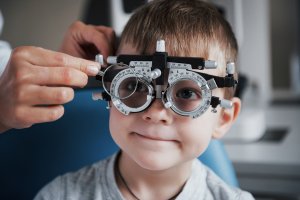
Q: How is an eye examination given to an infant or young child?
A: If glaucoma is suspected in a child under the age of four, it is often necessary to perform an examination with the child under anesthesia. Under anesthesia, the doctor is best able to test the child’s intraocular pressure (tonometry) and evaluate the angles or dimensions of the eye (gonioscopy). Gonioscopy assists the doctor in determining whether the eye is functioning properly: producing, circulating, and draining the fluid or aqueous humor within the eye.
Q: Should I allow my twelve year old participate in regular activities and sports, despite his glaucoma?
A: Your child’s participation level depends on his level of vision and the condition of his eyes. You should speak with your child’s ophthalmologist to determine what level of activity would be acceptable and what could be damaging. It is possible for a child to have a normal and active life if the glaucoma is under control and sight is good. If a child has had a corneal transplant, more caution is warranted and the child’s physical activity should not be strenuous or fast-paced.
Q: Is there a surgery that can cure childhood glaucoma?
A: Congenital, or infantile, glaucoma can often be treated through a goniotomy or trabeculotomy surgical procedure, although it may be necessary to have more than one operation. These procedures are invasive and intended to cut the trabecular meshwork (tear duct drain) in order to improve its drainage function. If these procedures are unsuccessful, it will become necessary to perform a trabeculectomy or implant an aqueous drainage device traditionally given to adults. If surgery is successful, it is sometimes considered a “cure” because the glaucoma may never become problematic or vision threatening, but it never really goes away. There is still a chance that conditions will surface years later, and that further treatment with medication and/or surgery will be required.
1-800-762-7132
Find a Doctor
Physician information including education, training, practice location and more.
Schedule an Appointment
Call 800-762-7132 or make an appointment online.